M. Pauling 1, B. Royston 1,
E. Sweeney 2, A. Douglas 1,
P.J. Howard 1.
(1) Merseyside and Cheshire Regional
Genetics Laboratories, Liverpool Women's Hospital, UK.
(2) Department of Clinical Genetics,
Alder Hey Hospital, Liverpool, UK.
Presented at the European Chromosome Conference, Vienna, July
1999
Case Report
After a chorionic Villus sample had revealed an apparently normal
female karyotype on direct preparation, this patient
was referred to this department at birth with bladder extrophy,
unusual streaked skin pigmentation and ambiguous
genitalia.
Cytogenetic Results
Peripheral Blood:
The peripheral blood sample at birth revealed an abnormal female
karyotype (fig 1)
46,XX,add(10)(p15). ish add(10)(wcp4+) de novo
The additional material on the short arm of chromosome 10 was shown
by Fluorescence In Situ Hybridisation (FISH) studies to be derived
from chromosome 4 material (fig 3), and it was deduced from the
banding pattern to most likely be 4q31.1 - 4qter. This anomaly therefore,
is a derivative chromosome 10 of the rearrangement t(4;10)(q31.1;p15).
As both parents were shown to have normal karyotypes this was considered
to be a 'de novo' event.
Fig 1. Peripheral blood karyotype:
46,XX,add(10)(p15). ish add(10)(wcp4+)
de novo.
(Click on image to enlarge)
Top of page
Skin Biopsies:
Biopsies taken from pigmented and non-pigmented areas of the patient's
skin showed the following Cytogenetic results
(confirmed by Molecular Cytogenetics-fig 3):
Pigmented Skin - 46,XX,idic(10)(qter-p15::p15-qter)/46,XX,der(10)t(4;10)
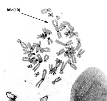
Non-Pigmented Skin - 46,XX, idic(10)(qter-p15::p15-qter)
As the anomalies seen in PB and skin were not detected prenatally
these are believed to be post Zygotic events.
Fig 2. Skin biopsy karyotype:
46,XX, idic(10)(qter-p15::p15-qter).
Click on an image to enlarge
Top of page
Possible Mechanism of Origin of Chromosome
Abnormality
It is postulated that this patient began embryonically with an apparently
normal female karyotype as detected in the Chorionic Villus sample
(CVS). A post zygotic mitotic anomaly lead to the formation of the
t(4;10) of which the der(4)t(4;10) was subsequently lost leaving
only the add(10)(p15) or der(10)t(4;10)(q31.1;p15) detectable in
the peripheral blood and in a mosaic form in the pigmented tissue.
This rearranged chromosome 10 then underwent a series of events,
the process being postulated as 'Uing' or U-type exchange first
described in 1989 by Wik Sjostedt et al (Hereditas 1989;111:115-123).
This U-type exchange event involves chromosome breakage, in this
case breakage at the translocated chromosome 4 end of the derived
chromosome 10 followed by a subsequent reunion between the sister<
chromatids. This event is thought to be generated by an abnormal
event in DNA replication. The result being the idic(10)(qter-p15::p15-qter)(fig
4) detected in both the pigmented skin in a mosaic form and in the
non-pigmented skin as the sole abnormality.
Fig 3. Mechanism for U-type exchange.
(Click on an image to enlarge)
Top of page
Skin Pigmentation
The pigmentary anomalies in this case have resulted directly from
the presence of a mosaic cell line. Numerous and diverse chromosomal
abnormalities have been reported in patients with pigmentary anomalies
distributed along the lines of Blaschko, which are linear areas
of skin differentiated from adjoining skin only by a difference
in pigmentation. They are aligned along the axis of limbs and circumferentially
around the trunk, and are thought to represent the tracts of migration
of melanocytes. Blaschko lines also show genetic mosaicism, characteristically
present in peripheral blood lymphocytes and/or skin fibroblasts
cultures. Chromosomal aberrations may conclude in abnormalities
of pigmentation as a result of the altered expression of genes in
the pigmentary pathway which map to a number of different chromosomes.
Our case confirms the relationship between chromosomal mosaicism
and anomalous skin pigmentation, particularly as lines of Blaschko
are followed. This stresses the importance in these cases of careful
evaluation of cells which should include a lymphocyte karyotype
and karyotypes from skin biopsies from both dark and light skin.
Fig 4. C-band image of idic(10).
Click on an image to enlarge
Conclusions
We postulate that in this case the pigmentary anomalies may have resulted
directly from the altered expression or gene dosage effect of specific
pigmentation genes or regulators of pigmentation localised on chromosome
10.
Top of page
|

